
Breast Imaging
The Specialist Breast Care Centre at Bon Secours Hospital Limerick offers a consultant‑led service delivered by highly experienced breast care experts. Patients receive comprehensive, empathetic, and truly patient‑focused care, with rapid access to both symptomatic and routine breast imaging and assessment when needed. Located within a state‑of‑the‑art healthcare facility, the Centre provides advanced diagnostics and cutting‑edge technology, supported by a strong commitment to national and international quality standards.
Specialist Breast Care Centre, Bon Secours Hospital Limerick
- Consultant led service by breast experts
- Comprehensive, empathetic, patient focused care
- Rapid access to symptomatic or routine breast imaging and assessment (if required)
- Advanced diagnostics & technology in a new, leading-edge healthcare facility
- National and International quality commitment
Having Routine or Symptomatic Breast Imaging
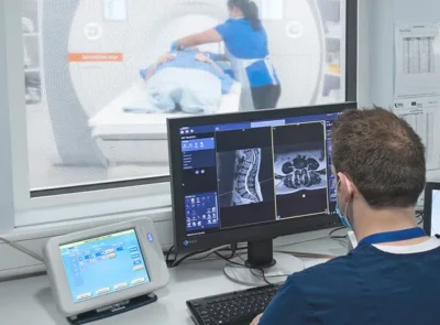
Our dedicated breast care service delivers thorough and compassionate care for patients with breast concerns/symptoms (symptomatic), and for those without symptoms (asymptomatic), within a modern, advanced facility with advanced imaging capabilities. Our expert team of consultant breast surgeons, radiologists, pathologists and oncologists, along with specialist nurses and radiographers, work together ensuring exceptional, coordinated care centred on patient needs. We provide our patients with prompt assessments, routine imaging and surgical options for breast cancer and other breast issues, in line with the highest quality assurance standards, both nationally and internationally.
- Professor Shona Tormey, Consultant Breast Surgeon
- Doctor Cressida Brennan, Consultant Radiologist
- Doctor Máire Lavelle, Consultant Pathologist
- Orla O’Neill, Clinical Nurse Specialist
- Lelanie Jansen, Clinical Specialist Radiographer
Breast cancer is a common cancer in Ireland. Almost 3,600 women and approximately 30 men are diagnosed with breast cancer each year. Breast cancer occurs when cells in your breast change and grow in an abnormal way. A tumour can occur if these cancer cells group together.
Early detection, however, makes breast cancer easier to treat and significantly improves the outcome.
Actions to increase your chance of detecting breast cancer earlier include:
- Attending routine breast screening appointments
- Being breast aware (self-examining the breasts for changes/symptoms)
- Visiting the GP promptly if symptoms arise
Although 9 out 10 patients with breast changes/symptoms receive a benign (non-cancerous finding), any breast changes require prompt investigation in a triple assessment clinic.
Examples of breast changes can include:
- A puckering or dimpling of the skin on the breast
- A lump in the breast or armpit
- A change in the skin around the nipple or discharge
- A rash on the breast area
- An unusual increase in the size of one breast
- One breast unusually lower than the other – nipples at different levels
- An enlargement of glands around the breast area
- An unusual swelling in the armpit
A GP referral is required to access the service. Your GP will decide on the best pathway of care for you based on your clinical risk.
Symptomatic – Triple Assessment Clinic
*We aim to offer our patients an appointment for triple assessment within one week of their visit to their GP*
- What is triple assessment?
In line with National Cancer Control Programme (NCCP) 2021 guidelines, the gold standard diagnostic approach for patients with symptoms (breast changes as outlined above) is a rapid access, diagnostic process combining three diagnostic tools in the same visit called Triple Assessment. These tools help maximise diagnostic accuracy, help detect early breast cancer and help rule out benign conditions, reducing unnecessary interventions, anxiety and worry.
Tests include:
- A clinical examination
- Diagnostic imaging – including a diagnostic mammogram and a breast ultrasound
- A biopsy if required
Our expert team compare all three parts of the triple assessment process to ensure the “Feel”, the “Picture” and the “Cells” all tell the same story.
- What is a clinical examination?
A clinical examination of the breast and armpit is performed by our consultant breast surgeon. The surgeon will gather the patient’s clinical history. Visual and palpable inspections are done to assess the area of concern. The outcome of the exam helps guide further imaging (the mammogram and ultrasound) to ensure the area of concern is appropriately visualised.
- What is a diagnostic mammogram?
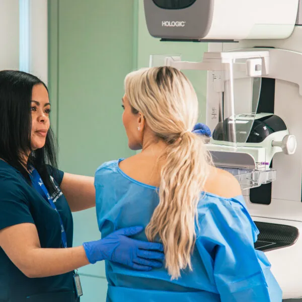
A diagnostic mammogram is a low dose X-ray of the breast. Our specialist radiographer positions your breast; applying compression to the breast once in the correct position. Compression is important to help visualise the tissues within the breast. Some patients may find compression uncomfortable, but it only lasts a few seconds. Typically, two projections of each breast are taken during a mammogram, but additional images may be taken if you have presented to the clinic with a symptom such as a lump. These images help plan the next steps in your care.
- What is a breast ultrasound?
A breast ultrasound is a painless imaging test performed by our consultant radiologist. It is used to evaluate breast changes, such as a new lump, in conjunction with a mammogram. Ultrasound uses high frequency sound waves to create real time pictures of the internal structures of the breast. It is particularly useful in determining whether a lump is a cancer or a benign (non cancerous) lump (i.e., cyst).
Ultrasound is typically, the first tool is accessing a symptom in a patient under 35 years of age where mammography can be less effective due to dense breast tissue.
- What is a biopsy?
A biopsy is the third and final check in triple assessment, also known as pathology. Many patients do not require a biopsy and for those who do, most biopsy results are benign (non cancerous).
A biopsy is performed by our consultant radiologist. It involves taking a tiny sample of cells from the area of the breast under review. Our consultant pathologist accesses these cells under a microscope.
The area is numbed prior to biopsy for patient comfort. Either X-ray or ultrasound is used to ensure the area of interest is sampled. Additional mammogram images may be taken after the procedure.
- Results pathway
Clinical exam and imaging (mammogram and ultrasound) only, results: same day
Clinical exam and imaging (mammogram and ultrasound) and biopsy/pathology – results: two weeks
Although most biopsy results are normal, for patients who receive a cancer diagnosis, supplementary tests including MRI and PETCT may be recommended to further assist in diagnosis. Our compassionate care team will design a patient centred care plan and support the patient during their journey.
Routine
High risk patients
High risk patients include those with known inherited risk factors including genetic mutations such as the BRCA gene, those with multiple family members with breast or ovarian cancer, a male relative with breast cancer or those with high doses of radiation to the chest at a young age. High risk patients account for a small percentage of the population. Most patients are average risk. Average risk means that a patient has no specific factors as mentioned above – making them more likely to develop breast cancer than the public.
A patient worried about their increased risk of breast cancer should discuss their concerns with their GP.
For patients with known high risk, we offer annual breast MRI (>25 years of age) and annual mammograms (>40 years of age) as required.
Average risk patients
- What is a routine mammogram (average risk)?
A routine mammogram involves a low dose X-ray of the breasts on a woman without symptoms. Typically two images of each breast is performed. Our specialist radiographer will position the breasts on the X-ray machine and apply compression. Compression is important in achieving a good quality mammogram. Some patients find compression uncomfortable; however, it only lasts a few seconds.
- Frequency of routine mammography
Routine mammography can be performed annually from the age of 40 – 50. Routine mammography is recommended biennially there after.
- Benefits versus risks with routine mammography
National and International guidelines are continuously updating as breast cancer trends change. There is currently an increase in breast cancer in women under 50 with studies advocating for earlier screening as a result. Routine mammography can allow for breast cancer to be detected two years prior to the patient having symptoms. Early diagnosis means a need for less invasive treatment and a better patient outcome.
Routine mammography can, however, result in false positives – resulting in unneeded recall for assessment for the patient (approximately 10% of women attending their first screening). This can cause unnecessary anxiety and worry. Routine mammograms can also over treat cancers that may not have caused issues for the patient. Although rare, interval cancers (cancers detected between screening appointments) can also occur.
At Bon Secours Limerick, we help to mitigate these risks by relying on an experienced, skilled team. We use state of the art equipment; continuously advancing with emerging clinical evidence.
- Results pathway
Results are sent to the patient’s GP two weeks after the screening mammogram.
- What happens if there is an abnormal result?
Patients recalled for further assessment are invited to our triple assessment clinic. Here they will have additional imaging performed and a clinical exam.
- Preparation
Our patients may be more comfortable in trousers and a top (rather than a dress) as top garments are required to be removed during exams. We also ask our patients to refrain from using deodorant and shimmer body oils/moisturisers pre attending our clinic as cosmetic particles can be visible on mammogram. Necklaces and large earrings are required to be removed prior mammography.
- Can I have somebody with me?
Yes, you are welcome to have somebody with you.
- Directions to the breast clinic
The Breast Clinic is located at Bon Secours Diagnostic Imaging Clinical Services Hub Towlerton Ballysimon Limerick V94892F.
- Parking
Parking is available at the Bon Secours Diagnostic Imaging Clinical Services Hub.
- What happens when I arrive at the Clinic?
Our dedicated team will check you in upon arrival to the Clinic.
- Can I access the service (particularly a screening mammogram) if I have had previous breast augmentation/lipofilling/surgery?
Yes. Your GP will communicate this information with our team, however, patients should inform a member of staff on arrival to ensure a tailored pathway to suit needs.
- How long will I be at the clinic?
Triple assessment clinic – 3 hours approximately
Screening mammogram – 45 mins approximately
- Health Insurance
Breast imaging is covered by the following health insurance providers including VHI, Level Health, Garda Medical Aid and POMAS.
Find a scan centre near you
We provide breast imaging and routine mammography in the following location in Ireland:
Book a Scan in Limerick | Diagnostic Imaging Limerick | Breast Imaging